Oh! The places you can Grow! Accessory Breast Tissue
Read Time | 10 minutes
Working in lactation, I don't think it's TMI to tell you that I have two nipples and two breasts.
The odds are high that you too only have two nipples and two breasts.
But a small population of my readers will have been blessed with more "junk in the proverbial boob trunk" (ignore me!) and will have at least one extra breast feature somewhere on their body.
Whether you have two or more breasts or none at all, I am so happy that you've taken the time to join me in talking about this interesting but not oft-discussed topic.
To understand how and why breasts may grow in places outside the 'typical' location (on the chest wall), it may help to understand how breasts develop in general.
Note: I use the word breast in this post for ease of reading as I am describing all 'typical' breast features such as nipple, areola, mammary gland, etc. I understand not everyone has breasts, so if you have a term you prefer other than breast (such as chest, for example), feel free to substitute that in where it may apply!
Mammary gland development happens soon after conception
When you were a tiny little embryo, possibly before your parent even knew they were pregnant, your mammary glands were already beginning to develop!
Breast development timeline
Between the 3rd and 4th week of embryonic development, the primitive milk streak appears. The milk streak is a line of ectodermal cells (outermost layer of cells) that extends from the groin to the axilla (armpit) on each side of the body.
Soon after, roughly the 5th week of development, the ectodermal cells thicken, forming the milk line, followed by a regression of the rest of the cells from the chest to the groin. The regression of the milk streak leaves breast tissue only where one might expect.
From 6 weeks onward, breast development continues, forming the mammary gland, ducts, nipple, areola, etc.
Your breast framework was already in place by the time you were born. Hormonal stimulation during puberty and your menstrual cycle further breast growth; however, most drastic breast development takes place during pregnancy.
Sometimes there's a hiccup
If the milk streak doesn't regress fully, the ectodermal tissue may continue to follow typical breast development. Depending on certain factors, one part of the breast development will continue and not others.
This leads to variations in the presentation of breast tissue along the milk line.
Polymastia
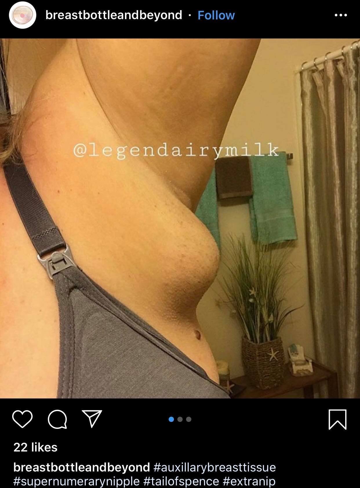
Polymastia, also referred to as hypermastia or accessory breast tissue, is the presence of extra breast tissue. The most common location of extra breast tissue is in the armpit.
If you have auxiliary accessory breast tissue, you may have noticed swelling or tenderness in your armpit during puberty, menstruation, or after delivery. In response to the decrease in progesterone, your armpit may have become engorged and painful a few days after delivery.
In most cases of auxiliary polymastia, there is breast tissue but no visible nipple or areola. But sometimes there are working ducts, in which case, you may have noticed the leaking of milk from your armpit.
Polythelia
Similar to polymastia, polythelia has many names- Hyperthelia, third nipple, supernumerary nipple.
It's easy to think of these two occurrences as distinct and separate, but in reality, it's just a spectrum, with different terms to describe different presentations.
As such, variations of additional breast tissue can be divided into 6 categories:
Category 1- Breast tissue with nipple and areola (Polymastia as described above)
Category 2: An extra nipple without areola but with breast tissue
Category 3: Breast tissue and areola but no nipple
Category 4: Breast tissue but no nipple or areola ( as described above)
Category 5 An extra nipple with an areola but underlying the external anatomy is fat rather than mammary gland (psuedomamma)
Category 6: An extra nipple with no breast tissue or areola (Polythelia)
Why are the categories important?
In regards to this blog post, the categories may be used as a reference to understand which variation is described in the following case studies.
Outside of this blog post, medical professionals love labels, and they use the category system to define the type of breast presentation for accessory breast tissue.
In the whole scheme of things, what category you may fall into is not super important, but it may be interesting!
Now for the super exciting part! Nearly all the case studies linked contain images.
Learning all the places, you can grow accessory breast tissue based on documented case studies!!
Vulva
Photo credit: Ed Uthman Modifications to original image by Shondra Mattos, IBCLC
A surprisingly common location of accessory breast tissue (comparatively), there are many case reports of documented vulvar ectopic breast tissue. The relatively high occurrence of breast tissue on the vulva is due to its location along the milk streak.
Five days post-delivery, a 29-year-old woman reported a painful swelling in her vulva. Following a diagnosis of accessory breast tissue without a prominent nipple or areola (polymastia), doctors determined that an open duct that would drain the breast was sutured after delivery. Removal of the sutures resulted in increase comfort for this patient.
As of 2012, 38 cases of ectopic breast tissue in the vulva have been reported. A 62-year-old post-menopausal woman sought care due to vaginal dryness and an uncomfortable lump, which turned out to be breast tissue in the vulva. A 45-year-old woman reported a lump in her vulva that had grown from the size of a peanut to the size of a lemon, after which she visited her health care provider. As you might imagine, biopsy showed it was normal breast tissue.
Vagina
Though accessory breast tissue along the milk line is most common, as we will see in this case, and others mentioned later, breast tissue can occur ANYWHERE on the body.
In the case of a 30-year-old woman from Korea, doctors identified a vaginal lump during the delivery of her son. Upon analysis, the composition of the lump was consistent with breast tissue.
Back
An 18-year-old female had surgery to remove the accessory breast tissue on her back, which had been present since birth. Not only did she have a large mass in the midline of her back, but she also had two fully formed and one underdeveloped nipple present on the extra breast.
A 15-year-old girl reported increasing pain in the swelling on her back, coinciding with breast development. Doctors observed accessory breasts in her interscapular region (between her shoulder blades) along with two nipples and seven areolas. Analysis of the excised tissue showed it was normal breast tissue typical with her stage of breast development.
Areola
Intraareolar polythelia, also known as nipple dichotomy, is an extremely rare presentation of polythelia in which two nipples are present on one areola.
A 24-year-old woman visited the clinic of a reconstructive surgeon due to a secondary nipple next to her right nipple. She reported that she had the extra nipple since she was 5-6 years old but that it had grown in the past couple of years. The plastic surgeons removed the extra nipple without complications.
Similarly, a 25-year-old woman reported an extra nipple next to her right nipple. She had the nipple since birth, and it doctors removed it without incident.
Thigh
Biologically female folk aren't the only ones affected by accessory breast tissue.
A 28-year-old male visited a surgical outpatient clinic due to a suspected lipoma (fat tumor) on the inner thigh of his left leg. Upon examination, doctors noticed a mass separate from his genitalia with a distinct areolar complex (areola and nipple) on the mass. The mass was excised in a manner similar to a full mastectomy, and the patient had an uncomplicated recovery.
A 64-year-old reported a nodule on the inner portion of her thigh, which she first noticed ten years prior and which caused friction with walking. Doctors visually diagnosed the nodule as an accessory nipple with accompanying breast tissue and offered a biopsy and excision, which she declined.
Forearm
A 15-year-old boy reported a soft mass on the hand side of his forearm. He explained that he first noticed the mass around 11 years old and noted that it originally excreted a thick white fluid when squeezed. The mass had grown from ages 11- 13, and at the time of examination, the drainage, while minimal, was thin.
The mass was removed, and analysis determined it was an accessory nipple.
Face
A 5-year-old boy from Turkey had a prominent nipple and visible areola on the left side of his head, between his temple and his left eye. The nipple, areola, and underlying tissue were removed and analyzed, and doctors determined it was a psuedomamma as there was no underlying breast tissue.
Are there any risks to having accessory breast tissue?
Having accessory breast tissue isn't inherently problematic, though it may cause discomfort or be a source of self- consciousness.
During lactation, accessory breast tissue that contains mammary tissue can leak if there are open ducts, and they can also develop mastitis.
The most significant concern, however, is the risk of breast cancer, which like the breast in the usual location, can affect breast tissue in the not-so-typical places.
The risk of breast cancer in unusual places is highlighted in this case study, in which a 70-year-old male was diagnosed with breast cancer of his perineum, specifically 'invasive breast carcinoma of no special specific type without evidence of nonneoplastic breast tissue.'
The risk of cancer is increased for those with accessory breast tissue, and it's for this reason that not only should any unusual lumps be biopsied and followed closely, but that many people opt to have the accessory tissue removed.
If you have accessory breast tissue and would like to share your experience to normalize it for others who may also be blessed with extra breast tissue, please do so in the comments below.
Additionally, you can join our growing community over on Facebook, where you're sure to get a lot of love for sharing!
Check out these other interesting blog posts


